With the advancement of early diagnostic methods, kidney cancer is increasingly being detected at earlier stages, often when tumors are less than 7 cm in diameter. For renal tumors measuring less than 4 or 7 cm, the most effective treatment approach involves a robotic-assisted technique that preserves healthy kidney tissue while excising the tumor. This procedure, known as Robot-Assisted Partial Nephrectomy (RAPN) or Robot-Assisted Nephron-Sparing Surgery, offers significant advantages. It maintains long-term cancer control without disadvantages and minimizes harm to the body, thereby effectively preserving kidney function.
In the majority of early-stage kidney tumors, a portion or a significant part of the tumor grows outward from the healthy kidney tissue. This is referred to as an exophytic renal tumor.
During robotic surgery, when a portion of the tumor is visible on the kidney’s external surface, the boundary between the tumor and the healthy kidney tissue can be readily identified. This clear delineation allows the surgeon to excise the tumor with precision, ensuring that no cancerous tissue remains at the surgical margin, thereby maintaining oncological safety. The robotic system’s enhanced visualization and dexterity facilitate the preservation of healthy renal parenchyma while achieving negative surgical margins.
What is an Endophytic Kidney Tumor?
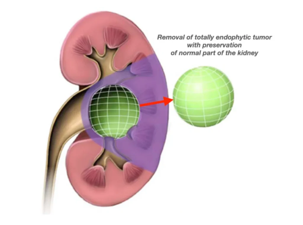
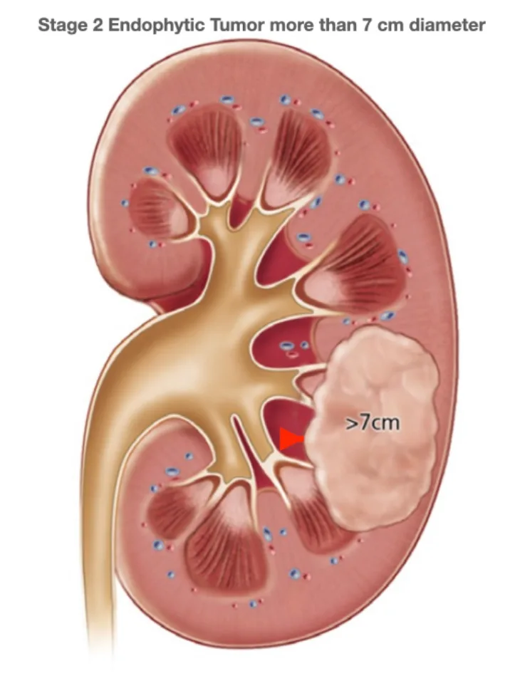
In some kidney tumors, the growth is entirely contained within the kidney and is not visible from the organ’s external surface. This type of tumor is known as an endophytic kidney tumor. Endophytic tumors are surrounded by normal renal parenchyma and do not protrude beyond the kidney’s outer contour. They can present surgical challenges due to their location and lack of external visibility.
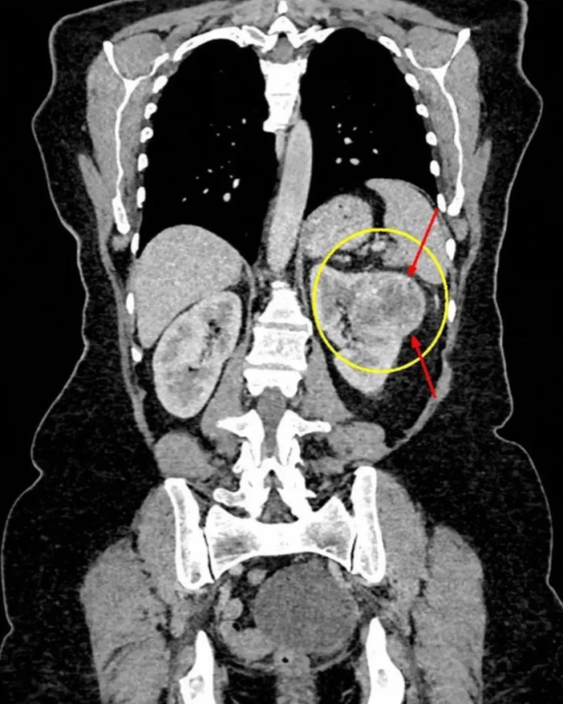
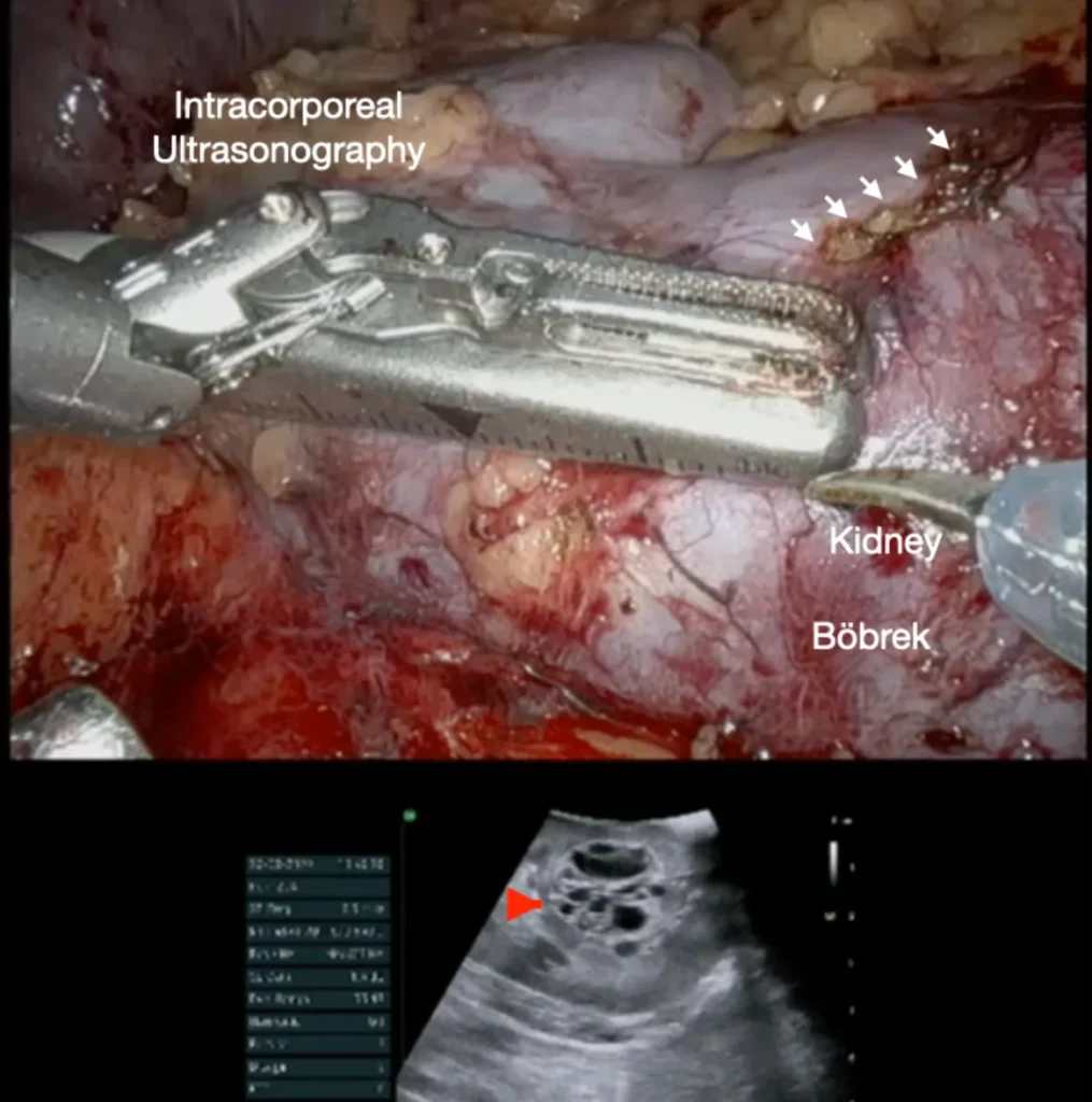
In cases where preoperative CT or MRI angiography has been performed, the tumor may still remain invisible from the kidney’s external surface during robotic surgery. Consequently, determining the precise location on the kidney’s surface to incise healthy tissue for tumor access and removal becomes challenging. Although attempts have been made to project MRI or CT images onto the robotic surgeon’s console screen using augmented reality techniques, these methods often fail to provide sufficient guidance. To overcome this limitation, a miniaturized sterile ultrasound probe, manipulated by the robotic arm, is introduced into the body during surgery. As depicted in the accompanying image, this approach enables clear visualization of even the most challenging endophytic tumors embedded within the kidney.
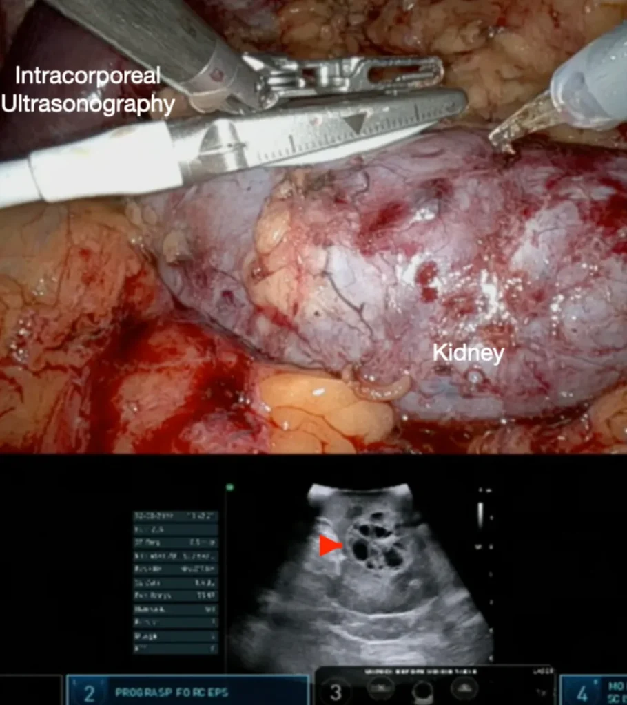
During robotic surgery, one robotic arm manipulates an ultrasound probe to delineate the boundaries of a tumor embedded within the kidney. Concurrently, another robotic arm, equipped with robotic scissors, is used to mark the tumor’s perimeter on the kidney’s surface. In the accompanying image, the red arrow indicates the tumor as visualized via ultrasound, while the white arrows highlight the kidney surface areas that have been delineated and marked during the robotic procedure. This technique allows for direct access to the endophytic tumor with minimal damage to healthy kidney tissue, facilitating the tumor’s removal while ensuring effective cancer control.
Even in cases where the embedded tumor is encased by major renal arteries or veins, Doppler ultrasonography can effectively delineate the tumor’s boundaries and its proximity to these blood vessels. This precise imaging allows for the tumor to be excised using robotic surgery with minimal damage to the kidney, ensuring oncological safety.
To observe the robotic ultrasonography-guided removal of a completely endophytic kidney cancer tumor with minimal bleeding, please click here or
visit YouTube Channel https://www.youtube.com/@tibeterdogru
Prof. Dr. Tibet Erdogru
Uro-Klinik Istanbul